Thursday 12 November 2015

24. Patent ductus arteriosus

If we go back to post 5 which deals with circulation in the unborn baby we will remember the 'ductus arteriosus' which is a very important structure in fetal life. It is through the ductus that blood circulates through the lower half of the baby. Usually the ductus arteriosus closes spontaneously after birth due to the effect of oxygen in the air that the baby breathes in. Closure of the ductus can take anywhere between 24-72 hours
In some babies the ductus does not close and can remain open resulting in Patent ductus arteriosus (PDA). If the PDA is small it may be of little consequence and sometimes can even be left alone. If however it is sizable it can lead to increased blood flow to the lungs which can cause breathlessness, poor feeding, sweating, poor weight gain etc in the baby. If left untreated in the long-term it can give rise to high pressure in the lungs (pulmonary hypertension) 

PDA is commonly found in premature babies and also in babies of mothers who have had Rubella infection. 

Timing of treatment depends on the size of the PDA. If the PDA is large it will need early treatment. If small it can be observed till 9-12 months of age giving time for spontaneous closure. PDA usually does not close spontaneously beyond the age of 1 year.

Treatment of PDA may be

(a) Medical: This is possible in preterm babies where certain drugs (NSAIDS like brufen or indomethacin) can be given to try and close the PDA. This has to be undertaken under the careful supervision of a children's doctor in hospital.


Image result for pda closure device
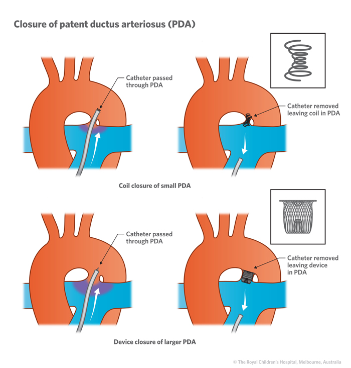
(b) Key hole: Here the PDA is closed using a button device. The device is introduced from the leg and inserted without open surgery across the PDA to close it. This technique may not be suitable for very larger PDAs in small babies.


Image result for pda closure device


(c) Surgery: This involves opening the chest and cutting and tying the PDA to eliminate it. This usually involves a longer hospital stay than key hole treatment.

Following successful elimination of the PDA the heart is essentially normal and we expect the patient to have a normal childhood and adult life.

Tuesday 21 July 2015

23. Specific Lesions: Double outlet right ventricle (DORV)

Double outlet right ventricle is a group of conditions where both the blood vessels i.e. the vessel to the body (aorta) and the vessel to the lungs (pulmonary artery) arise from the right pumping chamber. For a refresher on the normal heart please refer to Blog post number 4.

Double outlet right ventricle is not one condition but a group of conditions where the common feature is that both the vessels arise from the right pumping chamber (RV). It is almost always associated with a hole in the wall between the pumping chambers (Ventricular Septal Defect or VSD) and sometimes there is obstruction in the blood flow to the lungs (Pulmonary stenosis or PS).

DORV is divided into several types depending on the arrangement and position of the two blood vessels and also on the position of the VSD. Below is a diagram showing the various positions of the VSD in relation to the two blood vessels.

The position of the VSD and the presence of obstruction to lung blood flow will determine whether a child is breathless or not and also whether there is 'cyanosis' (Refer to Blog post No 6). Some babies with DORV will need early surgery as they are found to be very blue due to severe obstruction to lung blood flow. These patients will need an initial 'shunt' operation to improve lung flow and then maybe a year or so later they can have the complete operation. The aim with surgery in DORV is to try to bring the heart back to normal. This is achieved using many surgical techniques


BT Shunt operation                                                                         Complete Repair                                            


Image result for rastelli repairImage result for bt shunt


Sometimes it is not possible to perform complete repair and a staged approach has to be taken to separate the blue and red blood in the heart (Glenn operation at 6-12 months of age followed by Fontan operation at around 5-7 years of age)

Glenn Operation                                                                                       Fontan Operation


Image result for fontan operation


Following surgery there needs to be close follow up at regular intervals. There is sometimes a need for re-operation in these patients.

Thursday 2 July 2015

22. Specific lesions: Transposition of great arteries (TGA)

Hello after almost a year. 

In response to popular demand regarding information about specific defects of the heart I will aim to address each defect in a systematic fashion. I start today with a condition called Transposition of great arteries

This is a simple illustration of the condition.  To recap from one of the earlier posts:
The normal heart has 4 chambers, 2 on the right side and 2 on the left side.The right sided chambers contain oxygen poor or blue blood and the left sided chambers contain oxygen rich or red blood. The right and left sides of the heart are separated by a wall. ‘Blue’ blood from the right pumping chamber is pumped into the lungs through a vessel called the pulmonary artery. ‘Red’ blood from the left pumping chamber is pumped into the body through a vessel called the aorta.

In transposition of great arteries the two blood tubes are swapped across; the aorta arises from the right side and the pulmonary artery arises from the left side. This means that the blue blood from the right side circulates in the body resulting in a 'blue baby' and the red blood from the left side circulates in the lungs.

This condition is present right from the time the heart is formed at 6-8 weeks of pregnancy and can be detected during the ultrasound scan that is performed in early pregnancy. After birth the baby is noted to be blue as the oxygen levels in the blood are very low. If left untreated most of the babies with this condition will die within the first month of life and 95% will die within the first year of life. 

Sometimes these babies need urgent treatment after birth and a life saving balloon procedure may need to be performed (balloon atrial septostomy). This is done in order to improve the oxygen levels in the blood. This however is only a temporary procedure and the only definitive treatment is open heart surgery. This has to be carried out within the first few weeks of life and is called an Arterial Switch Operation. It involves swapping the blood vessels over so that they are connected to the correct side of the heart.


Although the operation carries a risk (success rate of around 90% in a good paediatric cardiac unit) the long term results of this procedure are very good and these patients go on to lead a near normal life.

If the child does not get prompt, timely treatment the risks of the operation increase. Also there is a point in time beyond which the arterial switch operation cannot be performed. In this case a less ideal operation called the atrial switch operation will need to be carried out.

In summary transposition of great arteries is a serious birth defect of the heart that needs early detection and timely treatment. Although the operation does carry some risk the long term outcome is generally very good and we expect most patients to lead a 'normal' life.

Please feel free to ask any questions that you might have.


Wednesday 30 April 2014

20. Pulmonary Hypertension patient support group

May 5th - World Pulmonary Hypertension day 

Pulmonary hypertension is a severe life limiting condition where the blood pressure in the lungs gets elevated due to thickening of the blood vessels within the lungs. This can be due to various causes; in some patients there is no identifiable cause and this is called idiopathic pulmonary hypertension; in others, it may be due to lung disease; the third group of patients is our focus as they have a form of pulmonary hypertension that is potentially preventable. These patients suffer from a condition called Eisenmenger syndrome. In this condition pulmonary hypertension is a preventable complication of a heart defect that is present from birth.  If the heart defect had been detected and treated early during childhood the pulmonary hypertension could have been prevented and the patient would have had a normal lifespan and quality of life.

Birth defects of the heart occur in approximately 1% of all babies born today.They can present at various ages depending on the type and severity of the defect. Some severe defects are obvious right from birth but other less severe conditions may be detected only later in life. Children with heart defects can have breathlessness and feeding difficulties. They are also more prone to getting chest infections and have slow weight gain.

Delayed detection and treatment of birth defects of the heart can lead to permanent damage and high pressure in the lungs (pulmonary hypertension) making the patient unsuitable for definitive treatment like surgery or keyhole therapy. This underlines the importance of early detection and treatment of birth defects of the heart.

Once a diagnosis of Eisenmenger syndrome is made many patients are simply sent away with 'nothing can be done'. Although a complete cure may not be possible for these patients they do require regular follow up, medication and lifestyle advice on issues like pregnancy, contraception, exercise etc.
   
Until recently good medical therapy for this group of patients was not available and they were condemned to a life of gradual deterioration and early demise. Now, there are a wide range of medications that are available that can improve the quality of life of these patients and prolong their survival. The medications do come at a cost (between 750 and 5000 INR per month depending on the drug that is used) but they have been shown to improve the exercise capacity and quality of life of these patients and are indirectly believed to improve their long-term survival as well. Prior to starting medical therapy patients may require a cardiac catheter test to establish clearly that there is no scope for curative treatment.

Apart from medications these patients need counseling regarding various issues in life. Pregnancy in women with severe pulmonary hypertension should be strongly discouraged as it can lead to deterioration and death. Good advice regarding safe and reliable contraception should be given. Mild exercise like walking and swimming can be carried out but patients should be advised not to push themselves or participate in a competitive fashion.

Above all this, patients with pulmonary hypertension are often adolescents and young adults who suddenly find themselves having to deal with the burden of living with a condition without a cure. The doctor's consultation should allow them to express their doubts and apprehensions and help them face life with positivity and courage.

 To this end we have established a Pulmonary Hypertension Patient Support Group, which will function out of the CHIME office at MIOT hospital. We welcome all patients, families and members of the lay public affected by this cause to get involved with our movement and make our vision of eradicating preventable pulmonary hypertension a reality. Our first meeting was held on the 12th of April. Parents and children, came from places like Erode,  Vellore, Arkonam, and Thoothukudi and from Suburbs like Chrompet, Kolathur, Injambakkam. They shared their experiences and gained strength from each other. Parents said they always thought they were alone in this but they realized after coming to this meeting that so many people have been affected with PH. 
  
As a society we should aim to eradicate preventable pulmonary hypertension by early detection and treatment of heart defects in children. This can only happen with increased health education and awareness amongst the lay public and medical fraternity. Patients with pulmonary hypertension need support from their families, peers and the medical community to face the challenges that lie ahead.

I invite you to be part of this effort. Please email me on drsangeethaviswanathan@gmail.com for further correspondence


.